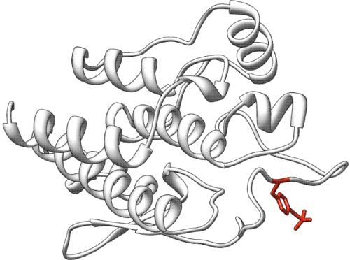
Predict the effect of somatic missense mutations on the activity and function of Serine/Threonine Kinase
Challenge: STK11
Variant data: registered users only
Last updated: 04 January 2022
This challenge is closed.
How to participate in CAGI6? Download data & submit predictions on Synapse
Make sure you understand our Data Use Agreement and Anonymity Policy
Summary
Serine/Threonine Kinase 11 (STK11) is considered a “master” kinase that functions as a tumor suppressor and nutrient sensor within a heterotrimeric complex with pseudo-kinase STRAD-alpha and structural protein MO25. Germline variants resulting in loss of STK11 define Peutz-Jaghers Syndrome (PJS), an autosomal dominant cancer predisposition syndrome marked by gastrointestinal hamartomas and freckling of the oral mucosa. Somatic loss of function variants, both nonsense and missense, occur in 15-30% of non-small cell lung adenocarcinomas, where they correlate clinically with insensitivity to anti-PD1 monoclonal antibody therapy. The challenge is to predict the impact on STK11 function for each missense variant in relation to wildtype STK11.
Background
Serine/Threonine Kinase 11 (STK11, NP_000446.1; NM_000455.5; GRCh38.p13 (GCF_000001405.39) chr 19, NC_000019.10 (1205778…1228431)) is a protein that functions as the catalytic subunit of a heterotrimeric complex comprised of STRAD-alpha and MO25 and is encoded by a 433 amino acid polypeptide. Germline STK11 loss of function defines Peutz-Jehgers Syndome (PJS) (OMIM #175200), an autosomal dominant cancer predisposition syndrome characterized by gastrointestinal hamartomas and hyperpigmented macules of the oral mucosa.
Recent clinical observations have correlated resistance to anti-PD1 monoclonal antibody therapy with Serine/Threonine Kinase 11 (STK11) LoF in lung adenocarcinomas driven by oncogenic KRAS variants (Koyama et al., 2016; Skoulidis et al., 2018). This genotype occurs in ~15% of non-small cell lung adenocarcinomas (NSCLCs) and is associated with a worse prognosis and increased rates of metastasis (Skoulidis et al., 2018; Calles et al., 2015; Ji et al., 2007; Nagaraj et al., 2017). STK11 is widely considered a “master kinase” and regulates numerous intracellular signaling networks impacting metabolism, proliferation and cell morphology (Hezel & Bardeesy, 2008). While most mammalian kinases are activated by autophosphorylation of their activation loop, the STK11 activation loop is stabilized in a conformation competent for substrate binding by interactions with MO25 (Zeqiraj et al., 2009). STK11 autophosphorylation occurs outside this loop in both the kinase (residues 49-309 and C-terminal regulatory (residues 309-433) domains (Sapkota et al., 2002; Baas et al., 2003), although the mechanisms linking autophosphorylation and STK11 activation remain poorly understood.
Somatically, STK11 variants represent the third most frequent genomic alteration in NSCLC, trailing only mutations in TP53 and KRAS (Sanchez-Cespedes et al., 2002; La Fleur et al., 2019). Known STK11 phosphorylation targets include 13 members of the microtubule affinity-regulating kinases family, AMPK being the most thoroughly studied (Lizcano et al., 2004; Nguyen et al., 2013). In response to nutrient stress, the STK11 complex phosphorylates and activates AMPK leading to inhibition of mTORC1-induced protein synthesis via phosphorylation of TSC2 and/or RAPTOR (Shackelford & Shaw, 2009), effectively inducing growth arrest. While the STK11/AMPK kinase cascade is undoubtedly important, a growing body of work reveals STK11 regulates myriad other biological processes and suggests the scope of STK11-loss-dependent regulation is vastly underappreciated (Esteve-Puig et al., 2014; Mehenni et al., 2005; Wang et al., 2014; Kline et al., 2013). An additional downstream target of STK11 activity includes the p53 signaling axis. STK11 has been shown to physically associate with p53 in the nucleus and activate p53-mediated transcriptional activity to regulate proliferation and apoptosis (Kaufmann et al., 2001; Zeng & Berger, 2006). The mechanism(s) governing STK11-dependent activation of p53 remain unclear and may occur directly via STK11-mediated phosphorylation of p53 on Ser15 and Ser392 (Zeng & Berger, 2006; Ma et al., 2018; Tiainen et al., 2002), or indirectly through the activation of AMPK and NUAK1 (Hou et al., 2011). In either case, p53 activation relies on intact STK11 kinase activity. If STK11 is absent or functionally impaired, p53-dependent transcriptional activity is reduced.
Experiment
As clinical decisions governing the use of anti-PD1 monoclonal antibody therapy for patients with KRAS-driven lung adenocarcinoma come to depend upon accurate STK11 functional status evaluation, the need for rigorous characterization of rare STK11 somatic variants grows urgent.
STK11, also known as LKB1, is an important tumor suppressor and energy sensor, regulating key signaling networks including the AMPK/mTOR axis (Lizcano et al., 2004; Nguyen et al., 2013; Shackelford & Shaw, 2009). Clinically, germline STK11 loss is pathognomonic for Peutz-Jeghers syndrome, an autosomal dominant cancer predisposition syndrome. Recently, STK11 functional status has emerged as a putative prognostic and therapeutic biomarker in patients diagnosed with KRAS-driven NSCLC (Skoulidis et al., 2018; Ji et al., 2007). Given 30,000 NSCLC patients per year are predicted to harbor this constellation of mutations, our ability to confidently assign functional deficits to novel somatic STK11 variants is paramount. Nonsense variants can generally be classified definitively as loss of function, especially when they occur early in the STK11 coding region. However, other somatic variant classes, including splice-site variants and missense variants, are more problematic. Nevertheless, accurately predicting the functional impact of splice-site and missense variants is essential to implementing personalized genomic medicine and currently remains an unmet need.
One of the current challenges we face is that there are many missense variants falling into the void of uncertain significance (VUS) whose contribution to disease is difficult to assess. Functional assays evaluating VUS can help provide the missing information, but may not capture all relevant variants, for example, variants resulting in the disruption of the STK11/STRAD-alpha/MO25 trimeric complex. Dr. Seward’s laboratory in the department of Pathology and Laboratory Medicine at the University of Vermont has functionally assessed the biologic impact, kinase activity and trimeric complex association of 28 missense mutations identified from primary NSCLC biopsy specimens at their institution.
STK11-dependent TP53-mediated transcriptional activation of a luciferase reporter
Plasmids containing cDNAs encoding each of the STK11 variants (STK11/eGFP) were transfected into A549 cells along with plasmids encoding the transfection control (PG13-luc) and the luciferase reporter (pRL-SV40) using Lipofectamine 3000 (Invitrogen, Carlsbad, CA) following manufacturer’s protocol. A549 cells lack STK11 but retain functional TP53.
Luciferase Assay
Eighteen hours post transfection, cell extracts were harvested per manufacturer’s recommendations using the Luciferase Assay Kit (Promega, Madison, WI). The luciferase assay was performed on a SpectraMax M4 Plate Reader (Molecular Devices, San Jose, CA). Three biologic replicates, each with three technical replicates, were evaluated by comparing the results for each variant with a WT STK11, and a known kinase dead point mutation (p.K78I). We found the results qualitatively binned with either the p.K78I or the WT. We did not observe intermediate phenotypes within the 28 variants evaluated.
STK11 putative autophosphorylation activity as measured by a gel-shift assay
Plasmids containing cDNAs encoding each of the mutant proteins fused to a FLAG-tag were transfected into A549 cells. This cell line lacks functional STK11 gene. STK11 heterotrimeric complexes (STK11/STRAD-alpha/MO25) were IP’d using anti-Flag beads and Kinase assays performed (+/- ATP, +/- lambda phosphatase). The kinase reactions were then subjected to SDS-PAGE electrophoresis and transferred to nitrocellulose membranes, followed by Western Blot analysis with anti-STK11 monoclonal antibody (E-9; #sc-374334, Santa Cruz Biotechnology; Dallas, TX, USA), and detected with anti-mouse-HRP purchased from Jackson ImmunoResearch Laboratories (West Grove, PA, USA). Again, the evaluated variants either demonstrated two distinct bands following blotting (an unmodified band and a shifted higher MW band, presumably the result of autophosphorylation, although the possibility of phosphorylation by another cell kinase cannot be excluded) indicating the variant behaved as WT, or only as single band (LoF), representing an inability to auto-phosphorylate. Addition of phosphatase eliminated the second band, confirming it was the product of phosphorylation. While the WT-like reactions didn’t result in a complete shift (the unmodified band was always detected), we failed to detect a shift (modified band) in those variants we classified as loss of function. In our hands the assay was essentially binary (one exception, where a weak shifted band was observed), the presence of a detectable modified band indicating WT-like activity.
Note: The results of the p53 mediated transcriptional activation assay and the putative auto-phorphorlyation assay were in agreement in 27/28 variants.
Prediction challenge
Participants are asked to submit predictions on the impact of the variants listed on STK11 function. The submitted prediction should be either “loss of function” or “wildtype”. Optionally, a comment on the basis of the prediction may be given if the submitter generates an intermediate or indeterminate result. In the previous challenges, it has been observed that predictions often cluster more with other predictions other than with the experimental value. Assessment will include metrics that recognize prediction sets that differ substantially from results provided by standard methods such as PolyPhen-2 and SIFT.
Prediction submission format
The prediction submission is a tab-delimited text file. Organizers provide a template file, which should be used for submission. In addition, a validation script is provided, and predictors should check the correctness of the format before submitting their predictions. In the submitted file, each row includes the following columns:
Each data row in the submitted file must include the following columns:
- AA substitution – The mutation found as listed in the prediction dataset file e.g., NP_000446.1:p.S31F. These variants are relative to UniProtKB Q15831 (STK11_HUMAN).
- Predicted activity – A non-negative prediction of variant STK11 function compared to wildtype STK11 on a scale where 0 = no activity, 1 = wildtype activity, >1 = higher activity than wildtype.
- Standard deviation – SD of the prediction in column 2, indicating confidence in prediction.
- Prediction (STK11 autophosphorylation) – A non-negative prediction of variant STK11 function compared to wildtype STK11 on a scale where 0 = no activity, 1 = wildtype activity.
- Standard deviation – SD of the prediction in column 4, indicating confidence
- Comment – Optional brief comment
In the template file, cells in columns 2-6 are marked with a "*". Submit your predictions by replacing the "*" with your value. No empty cells are allowed in the submission. You must enter a prediction and standard deviation for every mutant; if you are not confident in a prediction for a mutant, enter a large standard deviation for the prediction. Optionally, enter a brief comment on the basis of the prediction, otherwise, leave the "*" in these cells. Please make sure you follow the submission guidelines strictly. In addition, your submission must include a detailed description of the method used to make the predictions, similar to the style of the Methods section in a scientific article. This information will be submitted as a separate file.
File naming
CAGI allows submission of six models per team, of which model 1 is considered to be primary. You can upload predictions for each model multiple times; the last submission before deadline will be evaluated for each model.
Use the following format for your submissions: <teamname>_model_(1|2|3|4|5|6).(tsv|txt)
To include a description for your method(s), use the following filename: <teamname>_desc.*
Example: if your team’s name is “bestincagi” and you are submitting predictions for your model number 3, your filename should be bestincagi_model_3.txt.
Dataset citation
Donnelly LL, et al. Functional assessment of somatic STK11 variants identified in primary human non-small cell lung cancers. Carcinogenesis (2021) 42(12): 1428-1438. PubMed
References
Baas AF, et al. Activation of the tumour suppressor kinase LKB1 by the STE20-like pseudokinase STRAD. EMBO J (2003) 22(12): 3062-3072. PubMed
Calles A, et al. Immunohistochemical loss of LKB1 is a biomarker for more aggressive biology in KRAS-mutant lung adenocarcinoma. Clin Cancer Res (2015) 21(12): 2851-2860. PubMed
Esteve-Puig R, et al. A mouse model uncovers LKB1 as an UVB-induced DNA damage sensor mediating CDKN1A (p21WAF1/CIP1) degradation. PLoS Genet (2014) 10(10): e1004721. PubMed
Hezel AF, Bardeesy N. LKB1; linking cell structure and tumor suppression. Oncogene (2008) 27(55): 6908-6919. PubMed
Hou X, et al. A new role of NUAK1: directly phosphorylating p53 and regulating cell proliferation. Oncogene (2011) 30(26): 2933-2942. PubMed
Ji H, et al. LKB1 modulates lung cancer differentiation and metastasis. Nature (2007) 448(7155): 807-810. PubMed
Kaufmann WK, et al. Aberrant cell cycle checkpoint function in transformed hepatocytes and WB-F344 hepatic epithelial stem-like cells. Carcinogenesis (2001) 22(8): 1257-1269. PubMed
Kline ER, et al. LKB1 represses focal adhesion kinase (FAK) signaling via a FAK-LKB1 complex to regulate FAK site maturation and directional persistence. J Biol Chem (2013) 288(24): 17663-17674. PubMed
Koyama S, et al. STK11/LKB1 deficiency promotes neutrophil recruitment and proinflammatory cytokine production to suppress T-cell activity in the lung tumor microenvironment. Cancer Res (2016) 76(5): 999-1008. PubMed
La Fleur L, et al. Mutation patterns in a population-based non-small cell lung cancer cohort and prognostic impact of concomitant mutations in KRAS and TP53 or STK11. Lung Cancer (2019) 130: 50-58. PubMed
Lizcano JM, et al. LKB1 is a master kinase that activates 13 kinases of the AMPK subfamily, including MARK/PAR-1. EMBO J (2004) 23(4) 833-843. PubMed
Ma Q, et al. Liver kinase B1/adenosine monophosphate-activated protein kinase signaling axis induces p21/WAF1 expression in a p53-dependent manner. Oncol Lett (2018) 16(1): 1291-1297. PubMed
Mehenni H, et al. LKB1 interacts with and phosphorylates PTEN: a functional link between two proteins involved in cancer predisposing syndromes. Hum Mol Genet (2005) 14(15): 2209-2219. PubMed
Nagaraj AS, et al. Cell of origin links histotype spectrum to immune microenvironment diversity in non-small-cell lung cancer driven by mutant Kras and loss of Lkb1. Cell Rep (2017) 18(3): 673-684. PubMed
Nguyen HB, et al. LKB1 tumor suppressor regulates AMP kinase/mTOR-independent cell growth and proliferation via the phosphorylation of Yap. Oncogene (2013) 32(35): 4100-4109. PubMed
Sanchez-Cespedes M, et al. Inactivation of LKB1/STK11 is a common event in adenocarcinomas of the lung. Cancer Res (2002) 62(13): 3659-3662. PubMed
Sapkota GP, et al. Identification and characterization of four novel phosphorylation sites (Ser31, Ser325, Thr336 and Thr266) on LKB1/STK11, the protein kinase mutated in Peutz-Jeghers cancer syndrome. Biochem J (2002) 362(pt2): 481-490. PubMed
Shackelford DB, Shaw RJ. The LKB1-AMPK pathway: metabolism and growth control in tumour suppression. Nat Rev Cancer (2009) 9(8): 563-575. PubMed
Skoulidis F, et al. STK11/LKB1 mutations and PD-1 inhibitor resistance in KRAS-mutant lung adenocarcinoma. Cancer Discov (2018) 8(7): 822-835. PubMed
Tiainen M, et al. Growth arrest by the LKB1 tumor suppressor: induction of p21(WAF1/CIP1). Hum Mol Genet (2002) 11(13): 1497-1504. PubMed
Wang YQ, et al. Downregulation of LKB1 suppresses Stat3 activity to promote the proliferation of esophageal carcinoma cells. Mol Med Rep (2014) 9(6): 2400-2404. PubMed
Zeng PY & Berger SL. LKB1 is recruited to the p21/WAF1 promoter by p53 to mediate transcriptional activation. Cancer Res (2006) 66(22): 10701-10708. PubMed
Zeqiraj E, et al. Structure of the LKB1-STRAD-MO25 complex reveals an allosteric mechanism of kinase activation. Science (2009) 326(5960): 1707-1711. PubMed
Dataset provided by
David J. Seward, M.D., Ph.D., University of Vermont Department of Pathology, HSRF 206, 149 Beaumont Ave, Burlington VT, 05445.
Revision history
20 May 2021: challenge announced
08 June 2021: challenge opens
01 September 2021: challenge closed on August 31
04 January 2022: citation to the published experimental data added
